Translate الترجمة لأي لغة

السبت، 25 مارس 2017

فاطمة بنت قيس وقصة طلاقها وبيان أنه كان علي قاعدة الطلاق أولا ثم العدة ثم التسريح المستوحاة من سورة البقرة ثم نسخ في سورة الطلاق تبديلا بالعدة أولا ثم الطلاق ثم التفريق


فاطمة بنت قيس وقصة طلاقها وبيان أنه كان علي قاعدة الطلاق أولا ثم العدة ثم التسريح المستوحاة من سورة البقرة ثم نسخ في سورة الطلاق تبديلا بالعدة أولا ثم الطلاق ثم التفريق
319 -الحديث الثاني : عن فاطمة بنت قيس " أن أبا عمرو بن حفص طلقها ألبتة ، وهو غائب - وفي رواية : { طلقها ثلاثا - فأرسل إليها وكيله بشعير ، فسخطته . فقال : والله ما لك علينا من شيء : فجاءت رسول الله صلى الله عليه وسلم فذكرت ذلك له ، فقال : ليس لك عليه نفقة }.وفي لفظ : { ولا سكنى - فأمرها أن تعتد في بيت أم شريك ، ثم قال : تلك امرأة يغشاها أصحابي ، اعتدي عند ابن أم مكتوم . فإنه رجل أعمى ، تضعين ثيابك ، فإذا حللت فآذنيني . قالت : فلما حللت ذكرت له : أن معاوية بن أبي سفيان وأبا جهم خطباني ، فقال رسول الله صلى الله عليه وسلم : أما أبو جهم : فلا يضع عصاه عن عاتقه . وأما معاوية : فصعلوك لا مال له ، [ ص: 574 ] انكحي أسامة بن زيد ، فكرهته ثم قال : انكحي أسامة بن زيد ، فنكحته . فجعل الله فيه خيرا ، واغتبطت به } .
الحاشية رقم: 6
" وفي الحديث دليل على جواز ذكر الإنسان بما فيه عند النصيحة . ولا يكون[ص: 577 ]من الغيبة المحرمة وهذا أحد المواضع التي أبيحت فيها الغيبة لأجل المصلحة . و " العاتق " ما بين العنق والمنكب .
وفي الحديث : دليل على جواز استعمال مجاز المبالغة ، وجواز إطلاق مثل هذه العبارة ، فإن أبا جهم : لا بد وأن يضع عصاه حالة نومه وأكله ، وكذلك معاوية لا بد وأن يكون له ثوب يلبسه مثلا ، لكن اعتبر حال الغلبة ، وأهدر حال النادر واليسير . 
 
وهذا المجاز فيما قيل في أبي جهم : أظهر منه فيما قيل في معاوية ; لأن لنا أن نقول : إن لفظة " المال " انتقلت في العرف عن موضوعها الأصلي إلى ما له قدر من المملوكات ، أو ذلك مجاز شائع يتنزل منزلة النقل ، فلا يتناول الشيء اليسير جدا ، بخلاف ما قيل في أبي جهم . وقوله " انكحي أسامة بن زيد " فيه جواز نكاح القرشية للمولى . وكراهتها له : إما لكونه مولى ، أو لسواده ، و " اغتبطت " مفتوح التاء والباء وأبو جهم المذكور في الحديث : مفتوح الجيم ساكن الهاء ، وهو غير أبي الجهيم الذي في حديث التيمم .
قلت المدون: حادثة طلاق فاطمة بنت قيس تمت وأبو عمرو ابن حفص في غزوة الي اليمن مع علي ابن أبي طالب وكانت هذه الغزوة في أوائل العهد المدني أي حين كانت أحكام الطلاق تحت مظلة سورة البقرة والتي كانت قد تنزلت ابان العامين الأولين للهجرة وظلت مهيمنة علي المجتمع المسلم حتي تبدلت قواعدها بعد نزول سورة الطلاق التي نزلت في غضون العام الخامس الهجري تقريباً ،وكانت القاعدة في الطلاق المتأسسة علي تنزيل سورة البقرة هي :
طلاق أولا ثم عدة ،
ثم نسخ ماجاء في سورة البقرة تبديلاً بما جاء في سورة الطلاق:


وقد ورد علي ذلك بعض الأدلة التي احتواها نص رواية فاطمة بنت قيس * وفاطمة بنت قيس كما جاء في سير أعلم النبلاء :
[ص: 319] فاطمة بنت قيس الفهرية(رضي الله عنها) إحدى المهاجرات وأخت الضحاك . 
 
كانت تحت أبي عمرو بن حفص بن المغيرة المخزومي ، فطلقها ، فخطبها معاوية بن أبي سفيان ، وأبو جهم ، فنصحها رسول الله - صلى الله عليه وسلم - وأشار عليها بأسامة بن زيد ، فتزوجت به .
وهي التي روت حديث السكنى والنفقة للمطلقة بتة .
وهي التي روت قصة الجساسة .
حدث عنها : الشعبي ، وأبو سلمة بن عبد الرحمن ، وأبو بكر بن عبد الرحمن بن الحارث بن هشام ، وآخرون .
توفيت في خلافة معاوية وحديثها في الدواوين كلها .
قلت المدون [وقد تقدم أن أبا عمرو بن حفص هو زَوْج فاطمة؛  ومنهم مَنْ قَلبه، فقال فيه: أبو حفص بن عمرو بن المغيرة. 
وقد تقدم في القسم الأول على الصواب.).(مشهور بكنيته.)-الإصابة في تمييز الصحابة.(أَخرجه أَبو موسى مختصرًا وقال: أَوردوه في الأَسامي.)(أخرجه ابن منده، وأبو نعيم)
(قيل: أَبو حفص بن المغيرة.) (قيل: أبو أحمد)
(أُمه دُرَّة بنت خُزَاعيّ بن الحويرث الثقفي.)  أسد الغابة.
(روى محمد بن راشد، عن سلمة بن أبي سلمة، عن أبيه: أن حفص بن المغيرة طلق امرأته فاطمة بنت قيس، على عهد رسول الله صَلَّى الله عليه وسلم ثلاث تطليقات في كلمة واحدة. ورواه عبد الله بن محمد بن عقيل، عن جابر قال: طلق حفص بن المغيرة امرأته.) أسد الغابة.(بعثه رسول الله صَلَّى الله عليه وسلم مع عليِّ حين بَعَث عليًا إِلى اليمن، فطلق امرأَته فاطمة بنت قيس الفِهرِية هناك، وبعث إِليها بطلاقها، ثم مات هناك.
 
* وقيل: عاش بعد ذلك.
أَخبرنا فتيان بن أَحمد بن سَمنيَّة بإِسناده عن القَعْنَبي، عن مالك، عن عبد الله بن يزيد ــ مولى الأَسود بن سفيان ــ عن أَبي سلمة بن عبد الرحمن، عن فاطمة بنت قيس: أَن أَبا عمرو بن حفص طلقها البتة، وهو غائب. فأَرسل إِليها وكيلُه بشعير فَسَخِطَتْه، فقال: والله مالك علينا من شيء. فجاءَت رسولَ الله صَلَّى الله عليه وسلم، فذكرت ذلك له، فقال لها: "لَيْسَ لَكَ عَلَيْهِ نَفَقَةٌ". وأمرها أَن تَعتَدَّ في بيت أُم شَرِيك. ثم قال: "تِلْكَ امْرَأَةٌ يَغْشَاهَا أَصْحَابِي، اعْتَدِّي فِي بَيْتِ ابْنِ أُمِّ مَكْتُومٍ، فَإِنَّهُ رَجُلٌ أَعْمَى. تَضَعِيْنَ ثِيَابَكَ..." الحديث.(*) ومثله روى الزهري، عن أَبي سلمة، عن فاطمة، فقال: أَبو عمرو بن حفص.)أسد الغابة. (كان خرج مع عليّ إلى اليمن في عَهْدِ النبي صَلَّى الله عليه وسلم فمات هناك. ويقال: بل رجع إلى أَنْ شهد فتوح الشام.)الإصابة في تمييز الصحابة.
 
(أبو عمرو هذا هو الذي كلم عمر بن الخطاب رضي الله عنه وواجهه في عَزل خالد بن الوليد. ذكر النّسائي، قال:‏ أخبرنا إبراهيم بن يعقوب الجوزجاني، قال‏:‏ حدّثنا وهب بن زمعة، قال‏: حدّثنا عبد الله بن المبارك، عن سعيد بن يزيد، قال:‏ سمعت الحارث بن يزيد يحدِّث عن علي بن رباح، عن ناشرة بن سمي اليزنيّ، قال:‏ سمعت عمر بن الخطَّاب يقول يوم الجابية في حديث ذكره:‏ وأعتذر إليكم من خالد بن الوليد، فإني أمرته أَنْ يحبس هذا المال على ضعفة المهاجرين، فأعطاه ذا البأس وذا اليسار وذا الشّرف، فنزعته، وأثبت أبا عبيدة بن الجراح، فقال‏ أبو عمرو بن حفص بن المغيرة: والله لقد نزعْتَ غلامًا ـــ أو قال عاملًا ـــ استعمله رسولُ الله صَلَّى الله عليه وسلم، وغمدتَ سيفًا سلَّه الله، ووضعْتَ لواءً نصبه رسول الله صَلَّى الله عليه وسلم، ولقد قطعْتَ الرَّحم، وحسَدْتَ ابن العم، فقال عمر:‏ أما إنك قريب القرابة، حديث السّنّ، تغضب لابن عمك. قال إبراهيم بن يعقوب:‏ سألت أبا هشام المخزوميّ ـــ وكان علامة بأسمائهم ـــ عن اسم أبي عمرو هذا. فقال‏: اسمه أحمد. وذكر البخاريّ هذا الخبر في التّاريخ، عن عبدان، عن ابن المبارك بإسناده نحوه، وأخرجه فيمن لا يعرف اسمه من الكُنى المجردة عن الأسماء.)الاستيعاب في معرفة الأصحاب.(قال البَغَوِيُّ: سكن المدينة)الإصابة في تمييز الصحابة.
 
[قلت المدون]والدليل علي أن حادثة طلاقه لفاطمة بنت قيس هو ما أوردة صاحب كتاب الإصابة في تمييز الصحابة من أنه خرج مع عليّ إلى اليمن في عَهْدِ النبي صَلَّى الله عليه وسلم فمات هناك. ويقال: بل رجع إلى أَنْ شهد فتوح الشام.)الإصابة في تمييز الصحابة.وهذه الفترة الزمنية واكبت السنوات الأولي من الهجرة أيام البعوث والسرايا الي البلدان حول المدينة المنورة [أي تشريعات سورة البقرة فيما يخص موضوعنا]،والتي كانت تمهد لغزوة بدر الكبري،
 
وقد وافق النبي صلي الله عليه وسلم وكيل ابي عمرو ابن حفص عندما ذهب اليها بخبر أبي عم ابن حفص بطلاقه إياها وأَرسل إِليها وكيلُه بشعير فَسَخِطَتْه، فقال: والله مالك علينا من شيء. فجاءَت رسولَ الله صَلَّى الله عليه وسلم، فذكرت ذلك له، فقال لها: "لَيْسَ لَكَ عَلَيْهِ نَفَقَةٌ". وأمرها أَن تَعتَدَّ في بيت أُم شَرِيك. ثم قال: "تِلْكَ امْرَأَةٌ يَغْشَاهَا أَصْحَابِي، اعْتَدِّي فِي بَيْتِ ابْنِ أُمِّ مَكْتُومٍ، فَإِنَّهُ رَجُلٌ أَعْمَى. تَضَعِيْنَ ثِيَابَكَ..." الحديث وهكذا تأكد أن فاطمة طلقت علي تشريع سورة البقرة حيث كانت من تطلق وقتها تعد مطلقة ، وتخرؤج من بيتها (لا سكني) ولا ينفق عليها (لا نفقة)لأنها بداهة قد صارت أجنبية علي زوجها مطلقة وبناءا عليه فأنني ألخص مسار التشريعين في حادثة طلاق فاطمة بنت قيس لأنه طلاق ثلاث

أولا في مسار التطليق ثلاثا حسب تشريعات الطلاق في سورة البـــــــــــــــــــقرة(2هـ)

كانت القاعدة التشريعية المنبثق منها أحكام الطلاق في سورة االبقـــــــــــــــــــرة هي:

طلاق   يتبعه عــــــــــدة    ثم    تسريح
وعليه :
فالطلقة الأولي1 يتبعها عدة1 استبراء  ثم تسريح وللزوج حق الرد(وبعولتهن أحق بردهن في ذلك إن أرادا إصلاحا) ولا نفقة ولا سكني
والطلقة الثانية2 يتبعها عدة2 وتسريح وحق الرد لكن لا نفقة ولا سكني في عدتها
والطلقة3 يتبعها عدة3 وتسريح ولا حق للزوج بالرد إلا أن تنكح زوجاً غيره وتذوق العسيلة ثم يُطلقها وتسرح فللأول حق الرجعه_وفي هذه الثالثة لا نفقة ولا سكني.
وهو ما أقر به النبي صلي الله عليه وسلم.

يعني اختصاراً موجزا(لأمر فاطمة بنت قيس المتأسس علي مسار التشريع في سورة البقرة(المنزلة في عام 2 هجريتقريبا):
طلقة1يتبعها عدة1 __وللزوج حق الرًدَّه__ولا نفقة ولا سكني
طلقة2يتبعها عدة2__وللزوج حق الرًدَّه__ولا نفقة ولا سكني
طلقة3 يتبعها عدة3 __وليس للزوج حق الرًدَّه__ولا نفقة ولا سكني


ثانياً : وأما في مسار التطليق ثلاثا حسب تشريعات الطــــــلاق في سورة الطـــــــــــــــــــلاق(5هـ)
فصارت القاعدة التشريعية المنبثق منها أحكام الطلاق في سورة الطلـــــــــــــــلاق هي:

عــــــــــــدة الإحصاء يتبعها طـــــــلاق ثم فـــــراق 
وتفسيره كالآتي:
 
عدة 1 يتبعها طلقة1 ثم تفريق 
وانتهي من حسابات الزوجين مسمي الرجعة(لأنها في العدة زوجة)
ولها النفقة والسكني أثناء العدة (ولا تخرج من بيتها)
وشأنه معها شأن سائر الخطَّاب إذا طلقها مع نهاية العدة
 ____****____
 
عــــدة2 يتبعها طلقــــة2 ثم تفريــق 
وانتهي من حسابات الزوجين مسمي الرجعة
ولها النفقة والسكني أثناء العدة(لأنها في العدة زوجة) 

وشأنه معها شأن سائر الخطَّاب إذا طلقها مع نهاية العدة

 ____****____
 
عــــــــدة3 يتبعها طلقــــــة3 ثم تفريـــــق
^وانتهي من حسابات الزوجين مسمي الرجعة وهما في العدة
 ولها النفقة والسكني أثناء العدة ،
 ^وشأنه معها شأن الخطَّاب إذا طلق الثالثة،لكن بعد أن تنكح زوجا غيرة وتذوق عسيلته ثم يطلقها كما صار الأمر علي تشريع سورة الطلاق(عدة أولا ثم طلاق ثم تفريق)

إلا أنه في التطليقة الثالثة لا تحل له حتي تنكح زوجاً غيرة وتذوق عسيلته فإذا طلقها بنفس مسار التشريع في سورة الطلاق حلت للأول 
 
وللتوسع في الفرق بين:(اضغط هذا الرابط
نسخ تقديم الطلاق علي العدة في أحكام الطلاق بسورة البقرة )
 
أحكام الطلاق المنزلة في سورة البقرة2هـ تقريبا _أولا ثم تبديل مكان العدة وزمانها (نسخ مكان العدة وزمانها من صدر العدة الي دبر العدة) كما تنزل بعد في سورة الطلاق 5هـ 
 
                                 روابط  رائعــــــــــــة                               


واقرأ هذه الروابط
مدونة الطلاف للعدة *
المغزي من ألفاظ سورة الطلاق المستعملة في السورة
الطلاق للعدة شريعة الله الباقية الي يوم القيامة
نظام الطلاق في الاسلام هو الطلاق للعدة
أول كتاب الطلاق للعدة
*المغزي من ألفاظ سورة الطلاق في تشريعات الطلاق
نظام الطلاق في الإسلام هو الطلاق للعدة في سورة ا...
مصحف المدينة الازرق
سورة الطلاق - سورة 65 - عدد آياتها 12
نظام الطلاق في الاسلام هو الطلاق للعدة ***
*الطلاق للعدة مفصل جدا
الطلاق للعدة مفصل جدا
عدم احتساب التطليقة الخاطئة
تابع س وج حول مواضيع الطلاق*
كيف لم يفهم الناس تنزيل التشريع في سورة البقرة برغم وضوح فارق التنزيل الزمني بينها.وبين سورة الطلاق
اهدار الشرع للتطليقة الخاطئة وعدم الاعتداد بها *
قول ابن حزم في الطلاق والتعقيب عليه**
نسخ تقديم الطلاق علي العدة في أحكام الطلاق بسورة البقرة
مدونة القرآن آية آية
أول كتاب الطلاق للعدة
الطلاق للعدة مفصل جدا وزيادات مهمة بالصفحة


الحصبة الألمانية

 الحصبة الألمانية  
من ويكيبيديا، الموسوعة الحرة

طفل مصاب بالحصبة الألمانية
sssssssssssssssssssssssssssssssssssssssssssssssssssssssssssssssssssssssssss
الحصبة الألمانية أو الحميراء (Rubella) هي مرض مُعدي يسببه فيروس الحصبة الألمانية ولا يسبب للطفل إزعاجا أكثر من إزعاج الزكام، وتستمر فترة حضانة الفيروس بعد أن يدخل الجسم من 14 إلى 21 يوماً وعادة يكون 18 يوما ثم تبدأ أعراض المرض في الظهور مثل ارتفاع درجة حرارة الجسم خلال يومين الأولين وقد تنتفخ الغدد الواقعة خلف الأذنين وحول العنق ويظهر الطفح في اليوم الأول أو الثاني ويتكون من بقع مسطحة حمراء قانية في منطقة الوجه، ثم ينتشر بسرعة إلى كافة أنحاء الجسم وتزول كل الأعراض في اليوم الرابع والخامس. ويكون المصاب معديا في الفترة من أسبوع قبل ظهور الطفح إلى مدة أربع أيام بعد ظهوره، ويكتسب الشخص مناعة دائمة بعد شفائه من المرض. وتعد الحصبة الألمانية أقل انتشاراً من مرض الحصبة، وهي لا تسبب أي وباء، ولكنها تحمل خطر الإصابة بالتهاب الدماغ في حالات نادرة جداً، أما التأثير الأكثر حدوثاً عند البالغين الذين يصابون بالحصبة الألمانية فهو انتفاخ المفاصل وتصلبها. ولا يحتاج مرضى الحصبة الألمانية إلى علاج خاص، ولكن هذا المرض قد يؤثر على الجنين وهو في رحم والدته (كأن يولد الطفل مشوها / مصابا بمرض القلب / فقدان للنظر أو السمع / متأخرا في نموه العقلي والجسدي أو غير ذلك)، ولذلك يجب أن تتجنب المرأة الحامل الاتصال بشخص مصاب بالحصبة الألمانية لتجنب العدوى.

أسلوب العدوى والانتشار
تنتقل الحصبة الألماني عن طريق العدوى بواسطة الرذاذ (أي استنشاق هواء يحتوي على فيروسات المرض) من الشخص المصاب إلي الشخص السليم وذلك عن طريق إفرازات الجهاز التنفسي مثل العطس أو مخاط الأنف تحدث العدوى، وفي حالة الجنين تنتقل إليه العدوى عن طريق المشيمة من أمه المصابة.
الأعراض
أ- ارتفاع خفيف في درجة الحرارة، لا يزيد بأي حال من الأحوال عن 38.4 م◦.
ب- تضخم وضعف في العقدة الليمفاوية في قاعدة الجمجمة وخلف الأذن، ويبدأ تضخم الغدد الليمفاوية قبل الطفح الجلدي بيومٍ واحدٍ وهو من العلامات المميزة لهذا المرض.
ج- بقع حمراء بسيطة تظهر علي الوجه، الجسم ثم الأذرع والساق.
د- ألم في المفاصل.
ه- لا يستمر ارتفاع الحرارة ولا الطفح الجلدي أكثر من ثلاثة أيام، حيث ينتشر الطفح من الوجه إلى باقي الجسم ويختفي بسرعة خلال تلك الفترة، لذلك يسمى هذا المرض أحيانًا "حصبة الأيام الثلاثة"، أما تضخم الغدد فقد يستمر لمدة من 7 - 10 أيام.
إصابة الجنين بالحصبة الألمانية
((أ)) - عند إصابة الجنين بالحصبة الألمانية ينتشر الفيروس في الدم، وقد يكون من مضاعفاتِ ذك إصابة المشيمة، وبالتالي انتقال العدوى إلى الجنين وقد وجد أن نسبة حدوث ذلك تتراوح بين:
47% إذا حدثت العدوى خلال الأسابيع الأربعة الأولى من الحمل.
22% خلال الفترة من 5 - 8 أسابيع.
7% خلال الفترة من 9 - 12 أسبوعًا.
6% خلال الفترة من 13 - 16 أسبوعًا.
* ثم بعد ذلك يصبح الجنين في الأغلب في مأمن من خطر الإصابة بالحصبة الألمانية.
((ب)) - أما عن درجة الإصابة فإنها تختلف باختلاف الأجنة وباختلاف توقيت الإصابة أثناء الحمل ولا يمكن التنبؤ بنتائجها في كل حالة على حدة إلا أن ناتج الإصابة قد يكون أحد هذه الاحتمالات:
نجاة الجنين.
موت الجنين.
تشوه الجنين:
"أ" العمــى: تحدث إذا كانت العدوى خلال الأسابيع الستة الأولى من الحمل.
"ب" الصــم: تحدث إذا كانت العدوى خلال الأسبوع التاسع من الحمل.
"ج" تشوهات القلب: تحدث إذا كانت العدوى في الفترة من (5 - 10) أسابيع من الحمل.
"د" تشوهات الأسنان: تحدث إذا كانت العدوى في الفترة من (6 - 9) أسابيع من الحمل.
"ه" التخلف العقلي: تحدث إذا كانت العدوى في بداية الثلث الثاني من الحمل.
* بعض التشوهات السابقة قد لا تُكتشف بعد الولادة مباشرة مثل تشوهات الأسنان والتخلف العقلي والصمم التي قد يتأخر اكتشافها حتى عمر من 2 - 4 سنوات.
الوقاية
تكون الوقاية بأخذ المصل الخاص بالحصبة الألمانية ويوجد هذا المصل منفردًا (مصل ضد الحصبة الألمانية فقط) أو متحدًا مع مصل الحصبة المعتادة والتهاب الغدة النكافية وهذا الطعم يعرف بـ (MMR) ويُسمَّى ثلاثي الحصبة، وفي الحقيقة فإن هذا المصل المشترك في غاية الأهمية لعملية الإنجاب، وذلك للوقاية من الحصبة الألمانية بمخاطرها التي عرفناها وكذلك الوقاية من الحصبة المعتادة التي قد تؤدي إصابة المرأة بها أثناء الحمل إلى موت الجنين وكذلك الوقاية من التهابات الغدة النكافية التي قد يكون من مضاعفات الإصابة بها ضعف الإنجاب عند الذكور إذا حدث التهاب بالخصيتين حيث يؤدي إلى حدوث عقم مؤقت بنسبة 13% من الحالات المصابة، لكن العقم المطلق احتمالية نادرة. ويجب أن يعطى المصل كالتالي:
جرعة عند سن 12 - 15 شهرًا.
جرعة عند سن (4 - 6 سنوات) أو (11 - 12 سنة).
بعد البلوغ: يعطي للسيدات غير الحوامل مع مراعاة تأجيل الحمل لمدة سبعة أشهر بعد التطعيم كإجراء وقائي لحماية الجنين.
العلاج
طريقة علاج الحصبة الألماني لا تقلل من فترة الإصابة بالمرض أما بالنسبة للجنين، فلا يوجد علاج للحد من مضاعفات الحصبة فعلاج الجنين المصاب بالمرض في رحم الأم يعتمد علي مدى حالة الجنين بعد التعرض للمرض. إذا حدثت إصابة فعلية بالحصبة الألماني عند الطفل أو الشخص البالغ فيجب إتباع الآتي:
الراحة التامة في الفراش (حسب إرشادات الطبيب).
التأكد من إجراء فحوص طبية علي الأشخاص الذين يتعاملون مع الطفل بشكل يومي (حسب إرشادات الطبيب).
يصف الطبيب بعض العقاقير لخفض الحرارة أو تخفيف ألم المفاصل.
تاريخ المرض
في يوم 22 يناير ،2014، أعلنت منظمة الصحة العالمية خلو كولمبيا من الحصبة الألمانية لتصبح بذلك أول دولة لاتينية تتخلص من هذا المرض.في يوم 29 أبريل،2015 أصبحت منطقة الأمريكتين أول منطقة تابعة لمنظمة الصحة العالمية تتخلص من هذا المرض."

 (من ويكبيديا)

حمل تحقيقات حديث عبد الله بن عمر

الجمعة، 24 مارس 2017

ممممممممممـ

الفيديوهات الطبية (موقع) وThe QT interval

الفيديوهات الطبية (موقع)

The QT interval

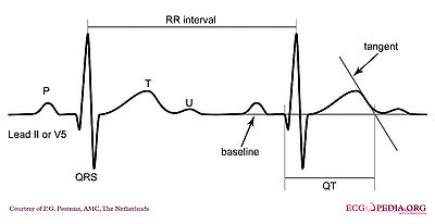
The QT interval starts at the onset of the Q wave and ends where the tangent line for the steepest part of the T wave intersects with the baseline of the ECG.
The eyeballing method to estimate QT prolongation. If the QT interval ends before the imaginary boundary halfway two QRS complexes, the QTc is probably normal. If the QTc reaches beyond the halfway line, the QTc is probably prolonged. This method is only 'valid' in registrations with normal (60-100/min) heart rates.
The normal QTc (corrected) interval The QT interval indicates how fast the ventricles are repolarized, becoming ready for a new cycle.
The normal value for QTc is: below 450ms for men and below 460ms for women as agreed upon by the ACC / HRS. [2]
In a recent ACC consensus document an expert writing group suggest that in a hospital setting the upper limit be raised to the 99th percentile of normal: 470ms in males and 480 ms in females, as approximately 10% to 20% of the general population have a QTc > 440m s. For both men and women QTc > 500ms is considered highly abnormal.[3]
If QTc is < 340ms short QT syndrome can be considered.
The QT interval comprises the QRS-complex, the ST-segment, and the T-wave. One difficultly of QT interpretation is that the QT interval gets shorter as the heart rate increases. This problem can be solved by correcting the QT time for heart rate using the Bazett formula: Formule QTc.png
Thus at a heart rate of 60 bpm, the RR interval is 1 second and the QTc equals QT/1. The QTc calculator can be used to easily calculate QTc from the QT and the heart rate or RR interval.
On modern ECG machines, the QTc is given. However, the machines are not always capable of making the correct determination of the end of the T wave. Therefore, it is important to check the QT time manually.
Alternatives to the Bazzett correction formula are the Fridericia, Framingham and Hodges formulas. The latter two perform better at high heart rates (>100 /min). [4][5]
  • Fridericia: QTc = QT{HR/60}1/3
  • Framingham: QTc = QT + 0.154{1 – (60/HR)}
  • Hodges: QTc = QT + 1.75 (heart rate - 60).
Although QT prolongation is potentially lethal, measurement of the QT interval by physicians is not standardized, since different definitions of the end of the T wave exist.[6] Most QT experts define the end of the T wave as the intersection of the steepest tangent line from the end of the T-wave with the base line of the ECG.[7] This leads to the following stepwise approach:
Stepwise approach to correct measurement of the QT interval
  1. Use lead II. Use lead V5 alternatively if lead II cannot be read.
  2. Draw a line through the baseline (preferably the PR segment)
  3. Draw a tangent against the steepest part of the end of the T wave. If the T wave has two positive deflections, the taller deflection should be chosen. If the T wave is biphasic, the end of the taller deflection should be chosen.
  4. The QT interval starts at the beginning of the QRS interval and ends where the tangent and baseline cross.
  5. If the QRS duration exceeds 120ms the amount surpassing 120ms should be deducted from the QT interval (i.e. QT=QT-(QRS width-120ms) ).
  6. Calculate QTc according to Bazett: Formule QTc.png. You can use the QTc calculator for this.
During ventricular pacing this method overestimates the QTc. The Framingham formula performs better during pacing, but still overestimates the QTc in sinus rhythm (in the same patient) by about 37-43 msec.[8]
In a pathological prolonged QT time, it takes longer than the normal amount of time for the myocardial cells to be ready for a new cycle. There is a possibility that some cells are not yet repolarized, but that a new cycle is already initiated. These cells are at risk for uncontrolled depolarization, induction of Torsade de Pointes and subsequent Ventricular Fibrillation.
Causes of QT prolongation
The QT interval is prolonged in congenital long QT syndrome, but QT prolongation can also occur be acquire as a results of:
  • Medication (anti-arrhythmics, tricyclic antidepressants, phenothiazedes, for a complete list see Torsades.org
  • Electrolyte imbalances
  • Ischemia.
QT prolongation is often treated with beta blockers.
If the QT segment is abnormal, it can be difficult to define the end of the T wave. Below are a number of examples that suggest how QT should be measured in these patients.
How to measure QT if the QT segement is abnormal
LastigeQT1.png
LastigeQT2.png
LastigeQT3.png
The T wave is broad, but the tangent crosses the baseline before the T wave joins the baseline. The QT interval would be overestimated when this last definition of the end of the T wave would be used.
The ECG does not meet the baseline after the end of the T wave. Still, the crossing of the tangent and baseline should be used for measurements.
A bifasic T wave. The tangent to the 'hump' with the largest amplitude is chosen. This can change from beat to beat, making it more important to average several measurements.

How do I begin to read an ECG?

How do I begin to read an ECG?   **    موقع الروابط

A short ECG registration of normal heart rhythm (sinus rhythm)
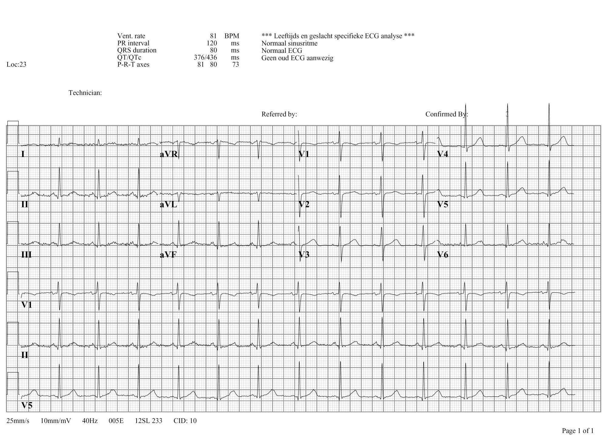
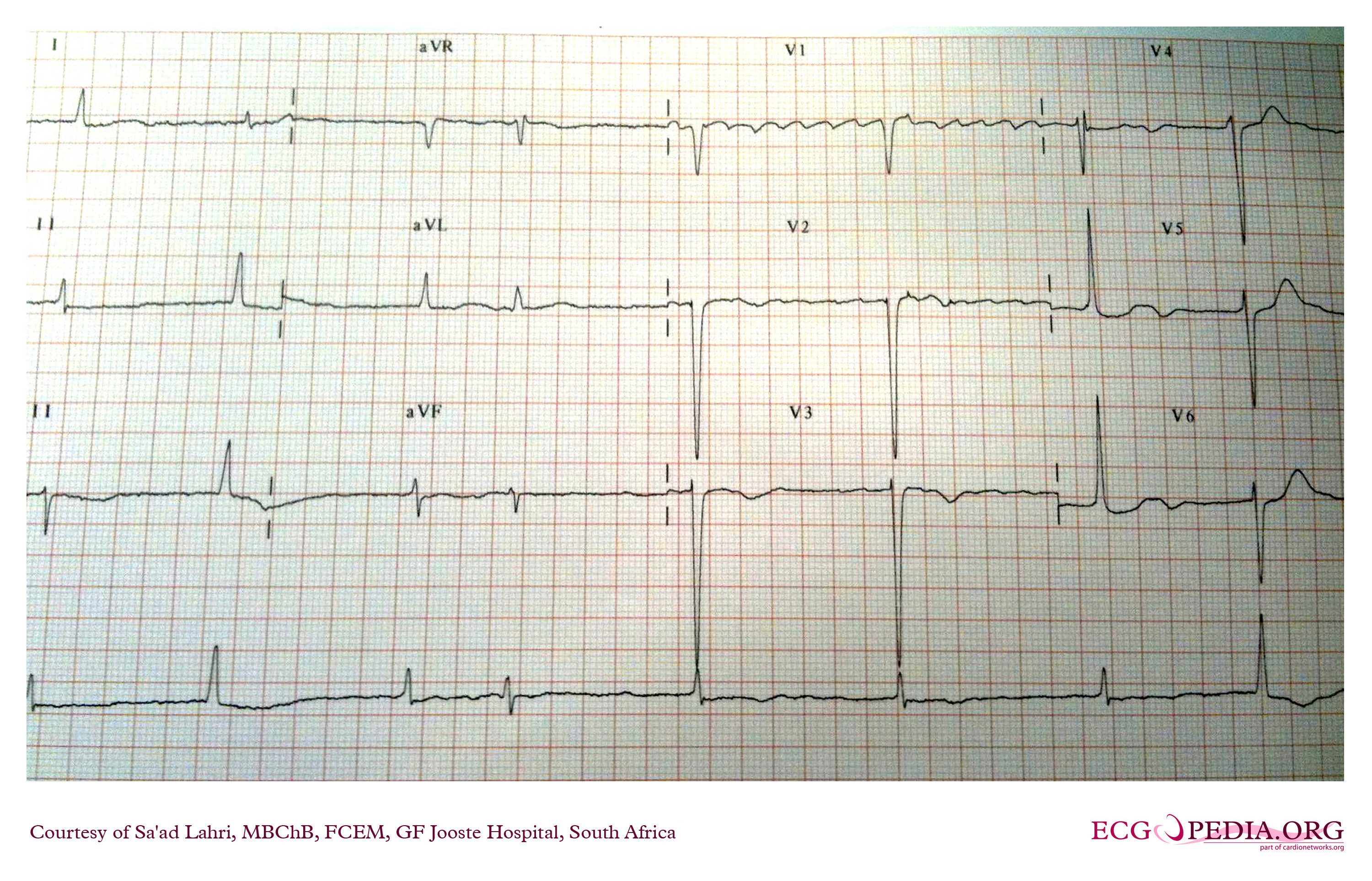
An example of a normal ECG. Click on the Image for an enlargement
Click on the ECG to see an enlargement. Where do you start when interpreting an ECG?
  • on the top left are the patient's information, name, sex and date of birth
  • at the right of that are below each other the Frequency, the conduction times (PQ,QRS,QT/QTc), and the heart axis (P-top axis, QRS axis and T-top axis)
  • further to the right is the interpretation of the ECG written (this often misses in a 'fresh' ECG, but later the interpretation of the cardiologist or computer will be added)
  • down left is the 'paper speed' (25 mm/s on the horizontal axis), the sensitivity (10mm/mV) and the filter's frequency (40Hz, filters noise from eg. lights)
  • finally there is a calibration on the ECG, on the beginning of every lead is a vertical block that shows with what amplitude a 1 mV signal is drawn. So the height and depth of these signals are a measurement for the voltage. If this is not the set at 10 mm, there is something wrong with the machine setting.
  • further we have the ECG leads themselves of course, these will be discussed below.
Note that the lay-out is different for every machine, but most machines will show the information above somewhere.


What does the ECG register?

The electrocardiogram
An electrocardiogram (ECG or EKG) is a register of the heart's electrical activity.
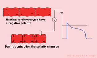
Just like skeletal muscles, heart muscles are electrically stimulated to contract. This stimulation is also called activation or excitation. Cardiac muscles are electrically charged at rest. The inside of the cell is negatively charged relative to the outside (resting potential). If the cardiac muscle cells are electrically stimulated, they depolarize (the resting potential changes from negative to positive) and contract. The electrical activity of a single cell can be registered as the action potential. As the electrical impulse spreads through the heart, the electrical field changes continually in size and direction. The ECG is a graph of these electrical cardiac signals.

The ECG represents the sum of the action potentials of millions of cardiomyocytes

Ion currents of the cardiomyocytes
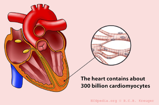
The heart consists of approximately 300 billion cells
In rest the heart cells are negatively charged. Through the depolarization by surrounding cells they become positively charged and they contract.
This movie shows the contraction of a single (rabbit) heart cell. The glass electrode measures the electrical current in the heart cell (with thepatch-clamp method). The electrical signal is written in blue and shows the action potential. Courtesy of Arie Verkerk and Antoni van Ginneken, AMC, Amsterdam, The Netherlands.

The individual action potentials of the individual cardiomyocytes are averaged. The final result, which is shown on the ECG, is actually the average of billions of microscopic electrical signals.
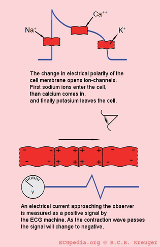
During the depolarization, sodium ions stream into the cell. Subsequently, the calcium ions stream into the cell. These calcium ions cause the actual muscular contraction.
Finally the potassium ions stream out of the cell. During repolarization the ion concentration returns to its precontraction state. On the ECG, an action potential wave coming toward the electrode is shown as a positive (upwards) signal. Here the ECG electrode is represented as an eye.


The electric discharge of the heart


موقع المشاركة


The electric discharge of the heart

The conduction system of the heart

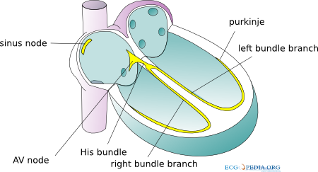
The sinoatrial node (SA node) contains the fastest physiological pacemaker cells of the heart; therefore, they determine the heart rate. First the atria depolarize and contract. After that the ventricles depolarize and contract. The electrical signal between the atria and the ventricles goes from the sinus node via the atria to the AV-node (atrioventricular transition) to the His bundle and subsequently to the right and left bundle branches, which end in a dense network of Purkinje fibers. The depolarization of the heart results in an electrical force which has a direction and magnitude; an electrical vector. This vector changes every millisecond of the depolarization. In the animation vectors for atrial depolarization, ventricular depolarization and ventricular repolarization are shown.

The different ECG waves

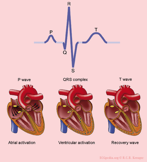
The origin of the different waves on the ECG
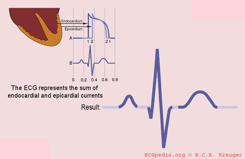
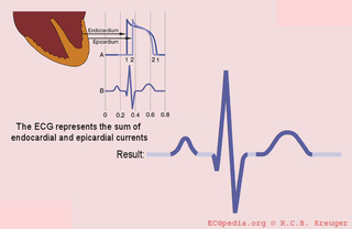
The QRS complex is formed by the sum of the electric avtivity of the inner (endocardial) and the outer (epicardial) cardiomyocytes
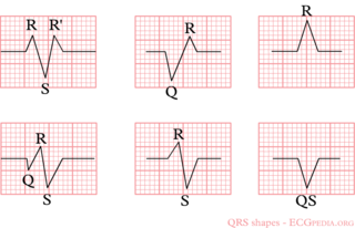
Example of the different QRS configurations
The P wave is the result of the atrial depolarization. This depolarization starts in the SA (sinoatrial) node. The signal produced by pacemaker cells in the SA node is conducted to the right and left atria. Normal atrial repolarization is not visible on the ECG (but can be visible during atrial infarction and pericarditis).
The QRS complex is the average of the depolarization waves of the inner (endocardial) and outer (epicardial) cardiomyocytes. As the endocardial cardiomyocytes depolarize slightly earlier than the outer layers, a typical QRS pattern occurs (figure).
The T wave represents the repolarization of the ventricles. There is no cardiac muscle activity during the T wave.
One heart beat consists of an atrial depolarization --> atrial contraction --> p-wave, ventricular depolarization --> ventricular contraction --> ORS-complex and the resting phase (including the repolarization during the T-wave) between two heart beats.
Have a look at this [animation of the heart cycle]
The origin of the U wave is unknown. This wave possibly results from "afterdepolarizations" of the ventricles.
The letters "Q", "R" and "S" are used to describe the QRS complex
  • Q: the first negative deflection after the p-wave. If the first deflection is not negative, the Q is absent.
  • R: the positive deflection
  • S: the negative deflection after the R-wave
  • Small print letters (q, r, s) are used to describe deflections of small amplitude. For example: qRS = small q, tall R, deep S.
  • R`: is used to describe a second R-wave (as in a right bundle branch block)
See figure for some examples of this.


The history of the ECG

A concise history of the ECG is presented in a different chapter.

The ECG electrodes

The limb leads
The chest leads
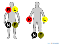
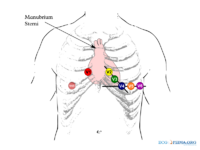
Electrical activity going through the heart can be measured by external (skin)electrodes. The electrocardiogram (ECG) registers these activities from electrodes which have been attached onto different places on the body. In total, twelve leads are calculated using ten electrodes.
The ten electrodes are:
  • The four extremity electrodes:
    • LA - left arm
    • RA - right arm
    • N - neutral, on the right leg (= electrical earth, or point zero, to which the electrical current is measured)
    • F - foot, on the left leg
It makes no difference whether the electrodes are attached proximal or distal on the extremities. However, it is best to be uniform in this. (eg. do not attach an electrode on the left shoulder and one on the right wrist).
  • The six chest electrodes:
    • V1 - placed in the 4th intercostal space, right of the sternum
    • V2 - placed in the 4th intercostal space, left of the sternum
    • V3 - placed between V2 and V4
    • V4 - placed 5th intercostal space in the nipple line. Official recommendations are to place V4 under the breast in women.[1]
    • V5 - placed between V4 and V6
    • V6 - placed in the midaxillary line on the same height as V4 (horizontal line from V4, so not necessarily in the 5th intercostal space)


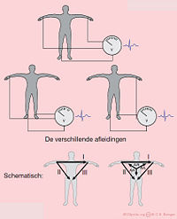
With the use of these 10 electrodes, 12 leads can be derived. There are 6 extremity leads and 6 precordial leads.

The Extremity Leads

The extremity leads are:
  • I from the right to the left arm
  • II from the right arm to the left leg
  • III from the left arm to the left leg
An easy rule to remember: lead I + lead III = lead II This is done with the use of the height or depth, independent of the wave (QRS, P of T). Example: if in lead I, the QrS complex is 3 mm in height and in lead III 9mm, the height of the QRS-complex in lead II is 12mm.
Other extremity leads are:
  • AVL points to the left arm
  • AVR points to the right arm
  • AVF points to the feet
The capital A stands for "augmented" and V for "voltage".
(aVR + aVL + aVF = 0)

The Chest Leads

The precordial, or chest leads, (V1,V2,V3,V4,V5 and V6) 'observe' the depolarization wave in the frontal plane.
Example: V1 is close to the right ventricle and the right atrium. Signals in these areas of the heart have the largest signal in this lead. V6 is the closest to the lateral wall of the left ventricle.

ECG variants

Besides the standard 12 lead ECG a couple of variants are in use:
  • The 3 channel ECG uses 3 or 4 ECG electrodes. Red is on the right, yellow on the left arm, green on the left leg ('sun shines on the grass') and black on the right leg. These basic leads yield enough information for rhythm-monitoring. For determination of ST elevation, these basic leads are inadequate as there is no lead that gives (ST) information about the anterior wall. ST changes registered during 3-4 channel ECG monitoring should prompt acquisition of a 12 lead ECG.
  • The 5 channel ECG uses 4 extremitiy leads and 1 precordial lead. This improves ST segment accuracy, but is still inferior to a 12 lead ECG. [2][3]
  • In vector electrocardiography the movement of electrical acitivity of the P, QRS and T wave is described. Additional X,Y and Z leads are recorded. Vector electrocardiography is rarely used nowadays, but is sometimes useful in a research setting.
  • In body surface mapping several arrays are used to accurately map the cardiac electrical wavefront as it moves over de body surface. With this information the electrical acitivity of the heart can be calculated. This is sometimes used in a research setting.

Color coding of the ECG leads

Two systems for ECG lead color coding are used: the AHA (American Heart Association) system and the IEC (International Electrotechnical Commission) system:

AHA (American Heart Association) IEC (International Electrotechnical Commission)
Location Inscription Colour Inscription Colour
Right Arm RA White R Red
Left Arm LA Black L Yellow
Right Leg RL Green N Black
Left Leg LL Red F Green
Chest V1 Brown/Red C1 White/Red
Chest V2 Brown/Yellow C2 White/Yellow
Chest V3 Brown/Green C3 White/Green
Chest V4 Brown/Blue C4 White/Brown
Chest V5 Brown/Orange C5 White/Black
Chest V6 Brown/Purple C6 White/Violet

Special Leads

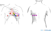
Leads V7,V8 and V9 can be helpful in the diagnosis of posterior myocardial infarction
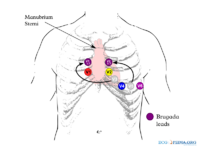
Changed lead positions of leads V3 and V5 to increase the sensitiviy to 'catch' a Brugada pattern on the ECG.
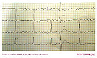
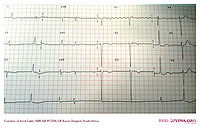
A patient with atrial fibrillation with a 'Lewis Lead' positioning of the leads. Compared with the normal lead configuration, the atrial signal is enlarged. Although some parts have a 'sawtooth' appearance consistent with atrial flutter, the rhythm is atrial fibrillation as there is a changing pattern in the atrial activity.
The same patient with a normal lead configuration. The rhythm is atrial fibrillation. The atrial activity in lead V1 is organized probably due to a organisation of electrical activity after it enters the right atrial appendage, close to lead V1.
Throughout history extra lead positions have been tried. Most are rarely used in practice, but they can deliver very valuable diagnostic clues in specific cases.
  • Leads to improve diagnosis in right ventricular en posterior infarction:
In case of an inferior wall infarct, extra leads may be used:
1. On a right-sided ECG, V1 and V2 remain on the same place. V3 to V6 are placed on the same place but mirrored on the chest. So V4 is in the middle of the right clavicle. The ECG should be marked as a Right-sided ECG. V4R (V4 but right sided) is a sensitive lead for diagnosing right ventricular infarctions.
2. Leads V7-V8-V9 can be used to diagnose a posterior infarct. After V6, leads are placed towards the back. See the chapter Ischemia for other ways of diagnosing posterior infarction.


  • Leads to improve detection of atrial rhyhtm:
In wide complex tachycardia, good detection of atrial rhythm and atrio-ventricular dissociation can be very helpful in the diagnosis process. An esophagal ECG electrode placed close to the atria can be helpful. Another, less invasive, method is the Lewis Lead. This is recorded by changing the limb electrodes, placing the right arm electrode in the second intercostal space and the left arm electrode in the fourth intercostal space, both to the right of the sternum. Furthermore gain is increased to 20mm/mV and paper speed to 50mm/sec.[4
  • Lead positioning to enhance detection of Brugada syndrome


Ladder diagram

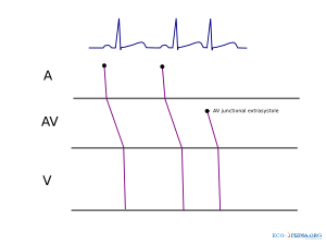
A ladder diagram is a diagram that shows the presumed origin of impulse formation and conduction in the heart. A = atrial, AV = AV node, V = ventricles
A ladder diagram is a diagram to explain arrhythmias. The figure shows a simple ladder diagram for normal sinus rhythm, followed by av-nodal extrasystole. The origin of impulse formation (sinus node for the first two beats and AV junction for the third beat) and the conduction in the heart are shown.
---------------------------

Technical Problems

Also read the chapter about Technical Problems. That will help you recognize electrical disturbances and lead reversals.


References

  1. Kligfield P, Gettes LS, Bailey JJ, Childers R, Deal BJ, Hancock EW, van Herpen G, Kors JA, Macfarlane P, Mirvis DM, Pahlm O, Rautaharju P, Wagner GS, American Heart Association Electrocardiography and Arrhythmias Committee, Council on Clinical Cardiology., American College of Cardiology Foundation., Heart Rhythm Society., Josephson M, Mason JW, Okin P, Surawicz B, and Wellens H. Recommendations for the standardization and interpretation of the electrocardiogram: part I: The electrocardiogram and its technology: a scientific statement from the American Heart Association Electrocardiography and Arrhythmias Committee, Council on Clinical Cardiology; the American College of Cardiology Foundation; and the Heart Rhythm Society: endorsed by the International Society for Computerized Electrocardiology. Circulation. 2007 Mar 13;115(10):1306-24. DOI:10.1161/CIRCULATIONAHA.106.180200 | PubMed ID:17322457 | HubMed [Kligfield]
  2. Rodrigues de Holanda-Miranda W, Furtado FM, Luciano PM, and Pazin-Filho A. Lewis lead enhances atrial activity detection in wide QRS tachycardia. J Emerg Med. 2012 Aug;43(2):e97-9. DOI:10.1016/j.jemermed.2009.08.057 | PubMed ID:20022196 | HubMed [Lewis1]
  3. Du Bois-Reymond, E. Untersuchungen über thierische Elektricität. Reimer, Berlin: 1848. [Dubois]
  4. Hoffa M, Ludwig C. 1850. Einige neue versuche uber herzbewegung. Zeitschrift Rationelle Medizin, 9: 107-144 [Hoffa]
  5. Waller AD. A demonstration on man of electromotive changes accompanying the heart's beat. J Physiol (London) 1887;8:229-234 [Waller]
  6. Einthoven W. Le telecardiogramme. Arch Int de Physiol 1906;4:132-164 [Einthoven]
  7. Einthoven W. Über die Form des menschlichen Electrocardiogramms. Pfügers Archiv maart 1895, pagina 101-123 [Einthoven2]
  8. Marey EJ. Des variations electriques des muscles et du couer en particulier etudies au moyen de l'electrometre de M Lippman. Compres Rendus Hebdomadaires des Seances de l'Acadamie des sciences 1876;82:975-977 [Marey]
  9. Márquez MF, Colín L, Guevara M, Iturralde P, and Hermosillo AG. Common electrocardiographic artifacts mimicking arrhythmias in ambulatory monitoring. Am Heart J. 2002 Aug;144(2):187-97. PubMed ID:12177632 | HubMed [Marquez]
  10. Hurst JW. Naming of the waves in the ECG, with a brief account of their genesis. Circulation. 1998 Nov 3;98(18):1937-42. PubMed ID:9799216 | HubMed [Hurst]
All Medline abstracts: PubMed | HubMed